Choosing Medical Infrared Imaging equipment requires more than comparing camera prices or promotional specifications. The right system must match the clinical purpose, patient population, examination environment, and professional workflow. Screening peripheral circulation, monitoring wound healing, and supporting musculoskeletal assessment may require different image quality and software capabilities.
Dr. John F. Ring, a recognized authority in medical thermography, described the method as “a non-invasive, non-contact, painless and radiation-free technique.” His statement highlights the technology’s practical value, but it should not be misunderstood. Medical infrared imaging records skin-surface temperature patterns. It does not independently confirm disease or replace clinical examination, laboratory testing, ultrasound, or other diagnostic methods.
Look closely at calibration procedures, thermal sensitivity, spatial resolution, temperature accuracy, lens options, and image-storage security. A camera used in a small clinic may need simple operation and rapid reporting. A research hospital may require advanced analytics, standardized protocols, and integration with electronic records. The room matters too. Drafts, direct sunlight, reflective surfaces, and recent exercise can distort thermal patterns. The details matter.
Manufacturers should provide transparent technical data, training resources, maintenance support, and evidence for intended applications. Regulatory status also deserves careful verification in the target country. A lower-cost device can appear attractive, yet poor calibration may create expensive clinical uncertainty. I would not pretend that one specification decides everything. Operator skill, patient preparation, and repeatable procedures often influence results just as strongly. The best choice is therefore not the most impressive camera, but the system that produces dependable information within a clearly defined clinical process.
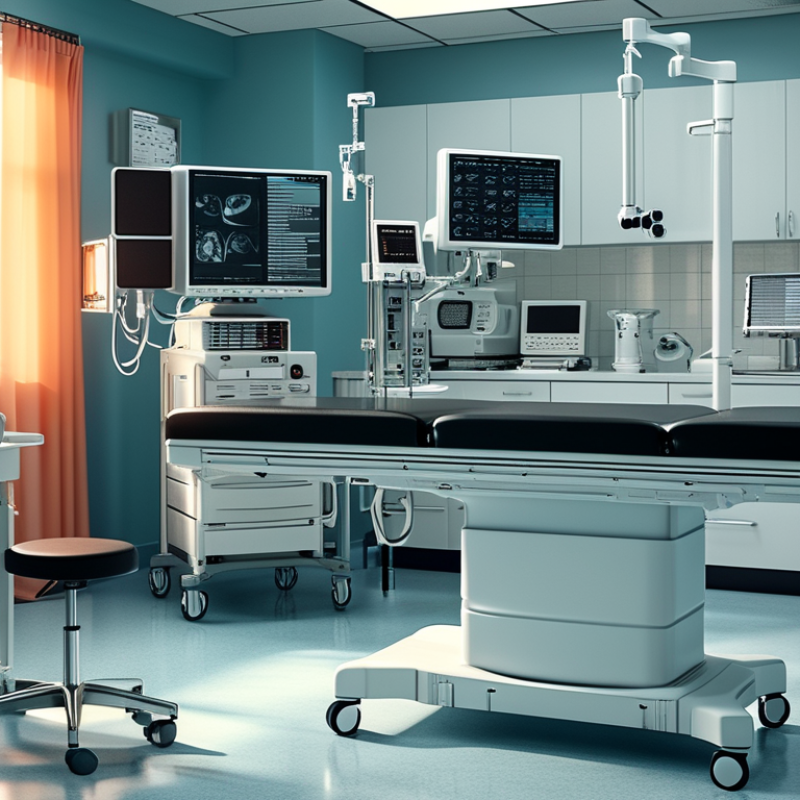
Before choosing medical infrared imaging equipment, define the clinical question precisely. Are you monitoring wound healing, assessing surface temperature, supporting vascular evaluation, or conducting research? Each purpose demands different imaging performance. A system for routine screening may prioritize fast capture and simple operation. A research workflow may require higher thermal sensitivity, adjustable temperature ranges, and detailed data export.
The target anatomy also matters. Small temperature differences can be clinically meaningful, but only when measurement conditions remain stable. Check spatial resolution, thermal sensitivity, calibration methods, frame rate, and the camera’s field of view. A wide view may help with body mapping, while close imaging can reveal localized changes. Room temperature, airflow, skin moisture, clothing, and recent exercise can affect results. Control them carefully. Do not ignore this.
Clinical integration deserves equal attention. Images should fit existing examination routines, patient records, and review procedures. Staff need practical training in patient positioning, distance, focus, and region-of-interest selection. Automated temperature alerts can save time, but they may also create distracting false positives. A warmer area is not automatically disease. Infrared imaging usually supports clinical judgment rather than replacing physical examination or established diagnostic tests. During evaluation, compare equipment performance with a trusted reference method and document repeatability across different operators. The first choice may be imperfect. Reassess it after real clinical use.
Choosing medical infrared imaging equipment requires more than comparing image sharpness. Different infrared technologies reveal different thermal information. Near-infrared systems can support surface observation under controlled lighting. Long-wave infrared cameras measure emitted heat from skin and nearby objects. Mid-wave systems may offer stronger sensitivity, but they often require more complex cooling and maintenance.
Sensor selection directly affects clinical usability. Uncooled microbolometers are compact, quiet, and practical for routine examinations. Cooled photon detectors can capture smaller temperature differences, especially during demanding measurements. However, greater sensitivity does not automatically mean better medical results. Check the noise equivalent temperature difference, spatial resolution, frame rate, and spectral response. A 320 × 240 sensor may miss a small fingertip region. A higher-resolution sensor may still perform poorly with unstable calibration.
Measurement capability deserves careful testing. Ask whether the system supports emissivity adjustment, reflected-temperature correction, and calibrated temperature readings. Skin, metal instruments, and glossy surfaces behave differently. A patient moving beside a cold window can distort the image. Room drafts can matter too. The measurement area should be documented consistently, including distance, angle, room temperature, and acclimatization time. Medical users should also verify calibration records and relevant safety documentation. A practical mistake is trusting colorful images too quickly. They look convincing. Yet image contrast is not the same as diagnostic accuracy. Test the equipment with known temperature references, repeat measurements, and review uncertain cases with qualified professionals.
How to Choose Medical Infrared Imaging Equipment?
Accuracy begins with repeatability, not attractive color maps. Check temperature resolution, calibration records, and the device’s stated measurement uncertainty. IEC 80601-2-59 sets requirements for screening thermographs, including performance verification and environmental control. A 2023 systematic review found wide variation in infrared imaging results across clinical studies. That variation should make buyers cautious. Ask for independent validation, not only manufacturer data. FDA guidance also warns that thermography should not replace proven diagnostic examinations.
Safety includes more than electrical protection. The World Health Organization reports that about one in ten patients experiences harm during healthcare, with over half considered preventable. Select equipment with clear alarms, secure data handling, and simple cleaning procedures. Confirm that operators can identify motion artifacts, cold rooms, wet skin, and poor positioning. These details can change a thermal image quickly.
Tips: Test the system in a real examination room. Measure setup time. Can one clinician adjust the camera, position the patient, and review images safely? Check whether handles support a steady wrist and whether the display remains readable under clinical lighting. Staff training matters, but complicated interfaces create avoidable mistakes. I have seen technically strong systems slow down a busy room. That is not a minor weakness. Record false alarms, repeated scans, and operator feedback during a trial period. A perfect specification sheet may still disappoint patients and staff.
| Evaluation Dimension | What to Evaluate | Practical Reference Point | Why It Matters | Verification Method |
|---|---|---|---|---|
| Intended Clinical Application | Confirm whether the system is intended for fever screening, physiologic monitoring, research, or adjunctive assessment. | The intended use, patient population, operating environment, and limitations should be clearly stated in the documentation. | Infrared images measure surface temperature and should not automatically be interpreted as a diagnosis of internal tissue temperature or disease. | Review the instructions for use, regulatory documentation, clinical evidence, and intended-use statement. |
| Temperature Measurement Accuracy | Check stated measurement accuracy, calibration conditions, target temperature range, and distance-to-target requirements. | For human-temperature screening systems, an accuracy target of approximately ±0.5°C is commonly used as a practical reference, subject to applicable regulations and validation requirements. | Small measurement errors can cause false alarms or missed elevated-temperature readings. | Test the equipment with a calibrated blackbody or certified temperature source under controlled conditions. |
| Thermal Sensitivity | Evaluate noise-equivalent temperature difference (NETD), detector stability, and the ability to distinguish small temperature variations. | A lower NETD indicates better thermal sensitivity; values at or below approximately 50 mK are commonly desirable for detailed surface-temperature assessment. | Higher sensitivity can improve visualization of subtle temperature differences, although it does not by itself establish diagnostic accuracy. | Compare manufacturer specifications and perform repeatability testing on a stable reference target. |
| Spatial Resolution and Field of View | Review detector resolution, lens angle, minimum measurement area, and the size of the anatomical region captured. | The selected field of view should allow the target area to occupy enough pixels for reliable region-of-interest measurement without excessive cropping. | Insufficient spatial detail can blend nearby regions with different temperatures and reduce measurement reliability. | Measure representative body areas at the planned working distance and inspect pixel coverage of the region of interest. |
| Environmental Control | Assess room-temperature stability, airflow, humidity, sunlight, reflective surfaces, and the subject's acclimatization time. | Use a controlled indoor area, avoid direct sunlight and drafts, and allow the subject to acclimatize according to the validated operating procedure. | External heat sources, perspiration, cosmetics, coverings, and recent physical activity can influence skin-surface temperature. | Create a site checklist and repeat measurements under normal clinical conditions before deployment. |
| Calibration and Quality Control | Check calibration interval, blackbody compatibility, automatic correction functions, and service requirements. | The system should have documented calibration procedures, traceable reference equipment, and a defined preventive-maintenance schedule. | Thermal sensors can drift over time or change performance with ambient-temperature variation. | Review calibration certificates, maintenance records, and quality-control results over time. |
| Patient and Operator Safety | Confirm that the equipment is non-contact, uses no ionizing radiation, and complies with relevant medical electrical safety requirements. | For clinical environments, review conformity with applicable requirements such as IEC 60601-1 and local regulatory rules. | Non-contact thermal imaging generally avoids exposure associated with ionizing imaging modalities, but electrical, optical, and data risks still require control. | Inspect safety certifications, risk-management files, electrical testing, and infection-control procedures. |
| Ergonomics | Evaluate weight, grip design, screen visibility, tripod or wall-mount options, cable placement, and adjustment controls. | Controls should be reachable without awkward wrist or shoulder positions, and the display should remain readable in the intended lighting conditions. | Good ergonomics reduce operator fatigue, accidental movement, and inconsistent positioning during repeated examinations. | Conduct a supervised trial with representative users and record setup time, posture, handling comfort, and repositioning frequency. |
| Ease of Use and Workflow | Assess startup time, focus or distance guidance, patient positioning prompts, region-of-interest tools, and alarm configuration. | A routine examination should require minimal manual steps and provide clear prompts for distance, positioning, and image acceptance. | Simpler workflows improve consistency and reduce training requirements and operator-dependent variation. | Run a timed workflow test with multiple operators and document error rates and training time. |
| Image and Data Management | Review image formats, metadata, user access, audit trails, encryption, export options, and integration with clinical systems. | Data handling should support applicable privacy requirements, role-based access, secure storage, and documented retention policies. | Thermal images may contain identifiable patient information and must be managed as clinical data where applicable. | Test export, backup, access-control, cybersecurity, and interoperability workflows before purchase. |
| Repeatability and Clinical Validation | Compare repeated measurements, different operators, different sessions, and relevant patient conditions. | Look for documented repeatability, reproducibility, sensitivity, specificity, and confidence intervals for the intended application. | A technically accurate sensor may still have limited clinical value if the workflow and clinical evidence are inadequate. | Review peer-reviewed evidence and conduct a local validation study using a defined reference method. |
| Maintenance and Total Cost | Include calibration, software updates, batteries, cleaning, accessories, training, service response, and replacement parts. | Calculate total cost of ownership over the expected service life rather than comparing purchase price alone. | Unplanned downtime and recurring calibration or software costs can significantly affect operational value. | Request a written service plan, maintenance schedule, warranty terms, and five-year cost estimate. |
Note: Performance targets should be confirmed against the equipment’s intended use, local regulatory requirements, applicable standards, and the healthcare facility’s validation protocol.
How to Choose Medical Infrared Imaging Equipment?
A medical infrared camera should fit the clinical workflow, not disrupt it. Observe how staff position patients, capture images, and review findings. The system should support quick acquisition without rushed examinations. Check whether images can move securely into the existing electronic health record or imaging archive. Manual file transfers create delays and increase identification errors. Small details matter, such as patient matching and timestamp accuracy.
Software deserves careful testing. Look for clear thermal scales, adjustable regions of interest, and consistent image annotations. Clinicians should compare current and previous examinations without searching through scattered folders. Useful software also records calibration status, operator details, and examination conditions. These fields strengthen interpretation and support later review. Ask for a live demonstration using realistic cases. A polished presentation can hide awkward daily steps.
Data management is not an afterthought. Confirm encryption, user permissions, audit trails, backup routines, and retention controls. The system should export standard formats when appropriate and preserve essential metadata. Test the process during network interruptions. It may still need a safe temporary storage method. Training should cover image capture, software updates, and troubleshooting. No workflow is perfect. A quick setup can encourage inconsistent positioning, while strict controls may slow busy clinics. Review both problems with users before purchase, then document practical acceptance criteria. A reliable evaluation includes technical staff, clinicians, and privacy personnel.
Compare the main procurement priorities before selecting a medical infrared imaging system. The weighting below emphasizes workflow integration, software capability, and secure data management.
How to use this chart: Evaluate each system against these four dimensions. Workflow integration covers patient identification, examination procedures, and reporting. Software includes image analysis, visualization, and export functions. Data management includes storage, access control, interoperability, and auditability. Clinical validation confirms that the equipment supports the intended medical use.
Compliance should be checked before image quality impresses the purchasing team. Confirm electrical safety, electromagnetic compatibility, and risk controls against IEC 60601-1 and ISO 14971. Ask for current certificates, test reports, software version records, and calibration procedures. A thermal camera may look accurate, yet drift after repeated cleaning or transport. Test it on a known-temperature reference source.
Support affects clinical continuity. Request documented response times, loaner equipment, remote diagnostics, and local repair capacity. The Ponemon Institute’s 2024 Cost of a Data Breach Report placed the average healthcare breach cost at $9.77 million. Therefore, verify encryption, access controls, audit logs, and update policies. Cybersecurity is part of equipment ownership, not an optional technical extra.
Calculate total cost over five years. Include training, calibration, batteries, software updates, accessories, downtime, and disposal. The U.S. Bureau of Labor Statistics projects strong growth for healthcare occupations through 2033, suggesting that staff time will remain expensive. Simple workflows can protect that scarce time. A few minutes saved per examination can matter. Do not trust a glossy return-on-investment spreadsheet. Compare real service records, warranty exclusions, and expected replacement cycles. The ECRI annual health technology guidance repeatedly emphasizes maintenance, cybersecurity, and lifecycle planning. I would also document weaknesses honestly, because uncertain data should not be treated as savings.
: Define the task clearly. You may monitor wound healing, surface temperature, vascular changes, or research data. Routine screening may need fast capture and simple controls. Research may require higher sensitivity, adjustable ranges, and detailed exports.
Check spatial resolution, thermal sensitivity, calibration, frame rate, and field of view. A wide view supports body mapping. Close imaging shows smaller regions. The best specification depends on the anatomy and clinical question.
Room temperature, airflow, moisture, clothing, and recent exercise can alter results. Keep conditions stable before capturing images. Allow the patient to rest when appropriate. Wet skin can mislead interpretation. Control the room carefully.
No. It usually supports clinical judgment and established diagnostic methods. A warmer region does not automatically indicate disease. Review motion artifacts, poor positioning, and environmental effects before interpreting findings.
Review calibration records, temperature resolution, and stated measurement uncertainty. Request independent validation, not only supplied performance data. Compare results with a trusted reference method. Repeat scans with different operators. The first choice may be imperfect.
Check secure data handling, clear alarms, cleaning procedures, and readable displays. One clinician should ideally position the patient and capture images safely. Test handles, wrist comfort, focus controls, and examination-room lighting. A technically strong device can still slow a busy clinic.
Observe real patient positioning, image capture, review, and reporting. Images should transfer securely into existing records or archives. Confirm accurate patient matching and timestamps. Test thermal scales, regions of interest, annotations, and comparison with earlier examinations. A polished demonstration can mislead.
Check encryption, user permissions, audit trails, backups, retention settings, and standard exports. Essential metadata should remain with each image. Test network interruptions and temporary storage procedures. Document practical acceptance criteria. Review problems with clinicians, technical staff, and privacy personnel.
Choosing the right Medical Infrared Imaging equipment begins with clearly defining its clinical purpose, such as screening, monitoring, diagnosis support, or research. Buyers should identify the required temperature range, image resolution, measurement distance, response speed, and environmental conditions before comparing infrared technologies and sensor types. The equipment should provide consistent, accurate results while supporting patient safety, comfortable operation, and efficient use by medical staff.
It is also important to assess how the system fits into existing clinical workflows. Consider software usability, image storage, reporting tools, data security, and compatibility with other systems. Compliance with applicable medical device requirements, documentation, training, technical support, maintenance, warranty coverage, and total ownership costs should be reviewed carefully. A reliable solution is not necessarily the one with the lowest purchase price; long-term value depends on dependable performance, manageable operating costs, flexible data management, and the ability to meet changing clinical needs.

Dianyang